Chapter 3 : The Forebrain and Somatosensory System

Brain: Contents Page

Sensations of Vibration and Proprioception (Kinaesthesia)


Vibration and Proprioceptive Senses: Receptors and Pathways

Proprioception, sometimes called Kinaesthesia, is the awareness of the relative positions of parts of the body, particularly the limbs. There is also an awareness of the degree of muscular effort necessary for movements to be executed.

Vibration Receptors in Muscle

Proprioceptors in the limbs include muscle spindles, Golgi tendon organs and joint receptors. Muscle spindles are very sensitive to vibration and can induce changes in muscle tone when vibratory stimuli are applied to a muscle,- the tonic stretch reflex.

Muscle spindles inform other neurons of the length of the muscle and the velocity of the stretch. This is particularly so for muscle that execute fine movements because muscle spindle density is highest in these muscle. There is also a high density of muscle spindles in the extensor muscle of the limbs that have to counter the effects of gravity and preserve posture.

Muscle Spindles are also very sensitive to vibration and monitor the position of small joints

Golgi Tendon Organs monitor the tensions at muscle-tendon junctions, and joint receptors sense the angle of each joint.

Vibration Receptors in Skin

Meissner's corpuscles also respond to vibration with an optimum frequence of around 30 Hz, whereas the highly sensitive Pacinian Corpuscles respond optimally to vibrations of around 250Hz. Sometimes the perception of vibratory stimuli is divided into two types on the basis of optimal frequency: frequencies of ~30Hz are referred to as 'flutter', and frequencies of ~200-300 are referred to as 'vibration'.

Pacinian Corpuscles are found in deeper tissues such as the deeper layers of the dermis and connective tissues. Vibration can also be sensed by muscle spindles, whose afferents also contribute to the dorsal column-medial lemniscal system.

All of these sensory receptors have myelinated axons.

*

The ways in which sensory receptors signal infomation is duscussed elsewhere

Dorsal Columns and Dorsal Column Nuclei.

Sensory modalities present in the dorsal column system are touch, pressure, vibration, and position sense. The sensory receptors involved are Pacinian Corpuscles, Meissner's Corpuscles and Mucsle Spindles. Position Sense (Kinaesthesia) involves afferent inputs form Muscles Spindles, Golgi Tendon Organs and Joint Receptors.

The large myelinated axons involved in the senses of vibration and kinaesthesia have cell bodies in the dorsal root ganglia. The afferent axons enter the dorsal horn and their main collaterals pass up the dorsal columns of the same side of the cord to reach the dorsal root ganglia.

The neurones of the dorsal column nuclei have axons that project through the medial lemniscus to the ventro-basal nuclei of the opposite side of the thalamus. These axons cross the midline within the medulla, and form the medial lemniscus, which passes through the pons and the dorsal tegmentum of the midbrain to reach the thalamus.

Somatosensory fibers are arranged in such a way as to preserve spatial information in their position, forming a map of the body surface known as a somatotopic map. Within this system, there are separate vibration and proprioceptive pathways (i.e.modality-specific private pathways) which reach the primary somatosensory cortex.

Wikipedia Commons

The Ventro-Basal Complex of the Thalamus   Top

Thalamic Nuclei concerned with Touch and Vibration

The medial lemniscus axons terminate in the ventrobasal complex of the thalamus, which consists of two main nuclei:

  • VPM is concerned with the head and receives inputs mainly from the trigeminal system.
  • VPL receives afferents from the rest of the body, and the leg is represented laterally within the nucleus, and arm medially.

Different areas ithin the nuclear complex retain their modality specificity. In other words, each sensory receptor type in peripheral nerves retain their characteristic properties in the post-synaptic neurones.

The nuclear complex has a central core dealing information concerned with light touch. Surrounding the core are a shell of neurones concerned with proprioception, and handling information from muscle spindles, Golgi Tendon Organs and joint receptors.

These nuclei project to the primary somatosensory cortex (SI, areas 3, 1, 2) in a modality specific and topographic manner, and pass through the internal capsule and corona radiata to reach the sensory cortex.

Representation of Vibratory and Proprioceptive Systems in the Somatosensory cortex   Top

Areas of Cerebral Cortex concerned with Vibration Sense

The primary somatosensory cortex (SI) is located in the post central gyrus, with the lower limb being represented on the medial surface of the hemisphere, and the head placed laterally near the Sylvian fissure. The pathways between sensory receptors and SI are modality specific and are anatomically and electrophysiologically distinct. As a result the somatosensory cortex is divided into strips, contain neurones that respond specifically to afferent inputs from

  • rapidly adapting skin receptors,
  • slowly adapting skin receptors,
  • muscle and tendon afferents, and
  • joint receptors
The columns of neurones within these strips are modality specific.

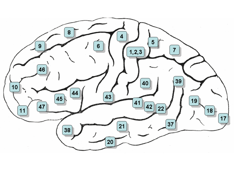
Brodmann's cytoarchitectonic map of the cerebral cortex was the result of an analysis of the size and shape of cortical neurones. There is a strong correlation between structure and function in the cortex, and the somatosensory cortex is in Brodmann's areas 1,2 and 3a + 3b. These represent parallel strips along the length of the post-central gyrus.

The different Brodmann areas within the post central gyrus are strips in parallel with the central sulcus, and the functional aspect of each strip is that it is modality specific - i.e. it handles information from only only one class of sensory receptor.

Pressure, flutter, and vibratory stimuli activate spatially distinct cortical columns in area 1. Area 3a handles information from muscle receptors; areas 3b and 1 handle the input from rapidly and slowly adapting receptors in skin, and columns of neurones within these areas preserve the modality specificity - i.e. there is no mixing of the modalities of sensory input within these columns.

In area 2, the input is from joint receptors: here the largest areas of cortex (number of cortical columns) are given to the position of large joints (hips, knees, etc) and smaller areas are concerned with smaller joints.

Top

Chapter 3 : The Forebrain and Somatosensory System

Brain: Contents Page

© HumanPhysiology.Academy 2014-2015